Oral exams are the foundation of lasting dental health. A thoughtful, thorough checkup does more than spot a cavity—it gives your dental team a complete, up-to-date picture of how your mouth is functioning and what it needs to remain healthy. By making oral exams a regular part of care, patients and clinicians can catch small problems early, personalize prevention strategies, and reduce the likelihood of more invasive treatment down the road.
At Corona Family Dental, we focus on practical, evidence-based exams that respect each patient’s comfort and concerns. During an exam we combine hands-on inspection with modern diagnostics and clear communication so you understand what we find and why it matters. This page explains what happens during an oral exam, why it’s important for whole-body health, and how imaging and cleanings fit into a proactive dental plan.
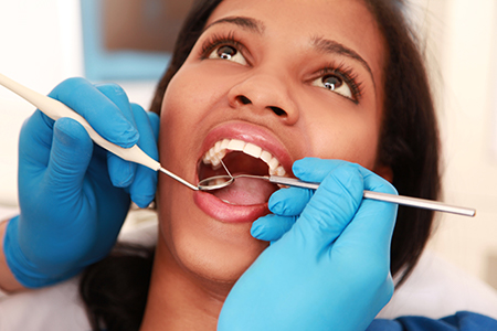
The first appointment with our team is designed to create a baseline: we document your dental history, review medications and medical conditions, and listen to your goals and concerns. That conversation guides a focused clinical exam that evaluates teeth, gums, bite, jaw joints, and the soft tissues of the mouth. Identifying current problems and potential risk factors helps us recommend sensible next steps tailored to you.
During the clinical portion of the exam we inspect for cavities, gum inflammation, wear patterns from clenching or grinding, and any unusual changes in the tissues. We also assess how your bite performs during normal function, which can reveal stresses that cause pain, fractures, or tooth movement over time. If needed, we test joint movement and listen for signs that the temporomandibular joint (TMJ) is not operating smoothly.
Diagnostic images are commonly part of a comprehensive first visit. X-rays or other scans let us see below the surface—roots, bone levels, and structures that aren’t visible to the naked eye. When imaging is recommended, we explain the type of image, what it shows, and how it informs the treatment plan so you can make informed choices about your care.
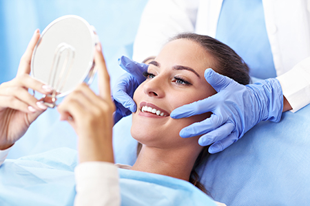
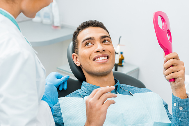
At the end of the exam we summarize findings and outline reasonable options for prevention or treatment, prioritizing steps that will protect your health with minimal disruption. Our goal is to create a clear, manageable plan and to answer any questions you have about risks, benefits, and expectations.
Oral tissues often reflect changes happening elsewhere in the body. During an exam we look for signs that might indicate systemic issues—persistent dry mouth, unusual ulcers, bleeding gums, or changes in tissue color and texture. Noticing these signs early can prompt timely medical follow-up and help you and your providers address root causes rather than only treating symptoms.
Researchers continue to explore links between oral inflammation and wider health concerns. Conditions such as cardiovascular disease, diabetes, and respiratory problems have been associated with chronic oral infections and gum disease. While an oral exam is not a medical diagnosis for these conditions, it can reveal warning signs that merit further investigation by your medical team.
Cardiovascular and circulatory concerns
Respiratory conditions that can be aggravated by oral bacteria
Blood sugar control issues such as diabetes
Gastrointestinal complaints that may relate to oral function
Neurological or cognitive associations under study
Conversely, medications and systemic diseases can create oral symptoms—dryness, tissue fragility, or delayed healing—that influence how we plan dental care. A thorough exam connects these dots so treatment recommendations consider your entire health profile, not just your teeth.
Routine checkups paired with professional cleanings are the most reliable way to keep oral disease at bay. Even diligent brushing and flossing at home can leave plaque in hard-to-reach areas; over time that plaque hardens into tartar, which requires professional instruments to remove. Removing these deposits reduces the bacteria that drive cavities and gum disease.
Regular visits also give us the chance to apply preventive measures and reinforce healthy habits. We’ll demonstrate techniques tailored to your mouth—whether you need tips for flossing around bridges, caring for orthodontic appliances, or modifying habits that contribute to wear. Education is a central part of each cleaning appointment because small changes at home lead to big improvements over time.
Frequency of visits depends on individual risk: many patients do well with twice-yearly exams and cleanings, while others benefit from more frequent monitoring due to gum disease, medical conditions, or increased risk of decay. We review your personal risk factors at each visit and recommend a schedule that balances convenience with the best protective care.
For children, routine visits also mean developmental monitoring. Early exams track tooth eruption and jaw growth so that any orthodontic concerns can be identified and managed at the right time, giving young smiles the best chance to develop naturally and healthily.
Imaging is an essential complement to visual inspection. Digital radiographs give clear, immediate views of tooth roots, bone levels, and hidden decay. Because the images are captured electronically, they reduce exposure times and provide faster, more efficient diagnostics compared with older film techniques.
Digital systems also streamline record keeping and collaboration. Images can be enlarged, adjusted for contrast, and stored in your electronic chart for side-by-side comparisons over time. That makes it easier to track slow changes and to communicate findings with specialists when advanced care is needed.
Beyond conventional 2D x-rays, three-dimensional imaging such as cone-beam computed tomography (CBCT) offers detailed views of bone anatomy and anatomical landmarks. When indicated, 3D scans improve diagnosis and treatment planning for complex cases, surgical procedures, and precise implant placement—always used judiciously and with patient safety in mind.
Because technology evolves quickly, we’ll discuss the benefits and limitations of any imaging we recommend so you understand how each image supports diagnosis and treatment decisions.
Different imaging types serve different purposes. Small intraoral images, like periapical films, show an individual tooth from crown to root and are ideal for spotting root issues or localized infections. Bitewing images focus on the crowns of posterior teeth and are especially useful for detecting early decay between teeth.
A full mouth series provides a comprehensive set of intraoral images when a broad survey of dental health is needed. Panoramic films capture both jaws in a single view, making them helpful for evaluating growth, impacted teeth, and overall jaw relationships. Cephalometric images—commonly used in orthodontics—offer a side profile view to assess skeletal relationships and plan orthodontic movement.
When precise three-dimensional detail is required, CBCT imaging provides volumetric views of bone and surrounding anatomy. This technology is invaluable for surgical planning, implant placement, and complex diagnostic situations where depth and spatial relationships matter.
We recommend imaging selectively based on each patient’s needs and the diagnostic questions at hand, balancing the value of information gained with a conservative approach to radiation exposure.
Regular oral exams connect preventive care, diagnostic imaging, and patient education into a coherent program that supports long-term oral health. If you have questions about what to expect at your next exam or which diagnostics may be right for you, please contact us for more information.
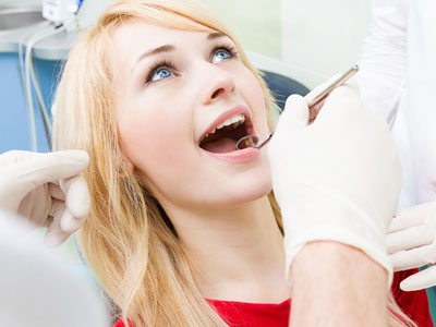
An oral exam begins with a review of your medical and dental history and a conversation about your symptoms, medications, and oral health goals. The clinician performs a systematic, hands-on inspection of teeth, gums, tongue, cheeks, and the soft tissues to identify decay, inflammation, or unusual changes. Bite and jaw function are evaluated to detect wear patterns, mobility, or signs of stress that can lead to pain or fractures over time.
When indicated, the exam includes diagnostic imaging such as digital radiographs to reveal problems below the surface, including root or bone changes. The dentist summarizes findings, explains how each observation relates to your oral and overall health, and outlines prioritized options for prevention or treatment. This structured approach helps build a clear, individualized care plan you can follow confidently at home and in future visits.
Oral tissues often reflect systemic health, so routine exams can reveal signs that warrant medical follow-up, such as persistent dry mouth, unusual ulcers, bleeding gums, or tissue changes. Researchers have identified associations between chronic oral inflammation and conditions like cardiovascular disease, diabetes, and respiratory complications, making oral screening a useful part of comprehensive health monitoring. While an oral exam does not diagnose non-dental diseases, it identifies warning signs that can prompt timely evaluation by your medical team.
An exam also considers how medications and systemic conditions affect oral healing, saliva production, and tissue resilience, which influences dental treatment decisions. By connecting oral findings with your broader health profile, clinicians create safer, more effective care plans that reduce the likelihood of complications. Early detection through regular exams frequently prevents minor problems from becoming complex, multi-disciplinary issues.
Before your first visit, gather a list of current medications, recent medical diagnoses, and any dental history you have, including past treatments or previous imaging. If you have specific symptoms—pain, swelling, or changes in sensation—note when they started and whether anything makes them better or worse. Arrive having brushed your teeth and avoid heavy food or tobacco use immediately before the appointment to make the exam and any imaging clearer.
If you are anxious or have special needs, let the office know when scheduling so staff can prepare appropriate accommodations and discuss sedation or relaxation techniques. Bring a photo ID and any referral information from other providers if applicable, and be ready to discuss your oral health goals so the clinician can tailor the exam to your priorities. Clear communication at the first appointment helps establish a baseline and a practical plan for ongoing care.
The most common interval for exams and professional cleanings is every six months, which works well for many patients to maintain oral health and catch early problems. Frequency is individualized based on risk factors such as gum disease, history of cavities, certain medical conditions, or medications that affect saliva; some patients benefit from more frequent visits, such as every three to four months. Your clinician reviews risk factors at each appointment and recommends a schedule that balances effective prevention with convenience.
Regular cleanings remove tartar that cannot be removed by home care alone and reduce the bacterial load that contributes to decay and periodontal disease. These visits also provide opportunities for targeted education—flossing techniques, appliance care, or habit modification—to reduce wear and improve long-term outcomes. Adhering to a tailored schedule is one of the best ways to prevent invasive treatment later on.
Digital imaging is a vital complement to clinical inspection, revealing root issues, bone levels, and decay that are not visible to the naked eye. Digital radiographs provide high-resolution images with lower radiation exposure and immediate availability, enabling faster diagnosis and clearer communication with patients and specialists. Electronic storage of images allows side-by-side comparisons over time, making it easier to track slow changes and evaluate the effectiveness of treatment.
Imaging choices are selected based on diagnostic need and patient safety; clinicians explain the type of image recommended and how it influences the treatment plan. Because images can be adjusted for contrast and magnified, they are a valuable tool for patient education and informed decision-making. Imaging is used judiciously, balancing diagnostic benefit with conservative radiation principles.
Cone‑beam computed tomography (CBCT) is recommended when three‑dimensional detail is necessary to diagnose complex conditions or plan precise surgical procedures, such as implant placement, assessment of impacted teeth, or evaluation of complicated anatomy. CBCT provides volumetric views of bone and adjacent structures that cannot be captured with standard two‑dimensional radiographs, improving accuracy in treatment planning for advanced cases. The decision to use CBCT is made selectively, only when the additional information will change the diagnosis or influence the care approach.
When CBCT is suggested, clinicians explain the specific diagnostic question the scan will answer, how it will influence treatment, and the steps taken to minimize radiation exposure. Images from CBCT are also useful for interdisciplinary communication with specialists when collaborative care is needed. This targeted use of advanced imaging helps ensure safe, predictable outcomes in complex dental care.
During an exam, clinicians assess gum health by measuring pocket depths, checking for bleeding on probing, and observing gum color, texture, and attachment to teeth. Visual inspection and periodontal measurements identify early signs of gingivitis and more advanced periodontitis so that treatment can begin before significant bone loss occurs. For decay, clinicians use visual and tactile examination combined with bitewing radiographs to detect interproximal lesions and early enamel breakdown that are not visible on the surface.
Detecting disease early allows for less invasive, more conservative treatments and targeted preventive strategies such as behavior modification, topical fluoride, or a focused hygiene plan. Periodic re-evaluation after treatment confirms healing and helps adjust maintenance frequency to prevent recurrence. Effective monitoring through exams and imaging reduces the risk of progressive disease and tooth loss.
An oral exam evaluates jaw movement, joint sounds, palpation tenderness, and muscle function to identify signs of temporomandibular joint (TMJ) disorders or muscle-related pain. Clinicians observe how the bite performs during function and assess for wear patterns, fractures, or tooth mobility that can indicate parafunctional habits such as clenching or grinding. If needed, the exam may include simple functional tests and documentation of symptoms to guide conservative management options.
Treatment often begins with noninvasive approaches like occlusal guards, behavior modification, physical therapy exercises, and short-term pain management while monitoring response to care. When imaging or specialist input is required, clinicians explain the purpose of further diagnostics and coordinate referrals as appropriate. The goal is to reduce pain and improve function through a staged, evidence-based plan that emphasizes conservative care first.
Pediatric oral exams focus on tooth eruption, bite development, and early detection of decay or habits that can affect growth, such as thumb sucking or tongue thrusting. Clinicians assess oral development at each visit, monitor jaw growth, and identify any orthodontic concerns that may benefit from early interceptive treatment. Examinations also include age‑appropriate hygiene instruction and preventive measures tailored to a child’s needs, including topical fluoride or sealants when indicated.
Early visits establish a positive relationship with dental care and give parents guidance on home care, diet, and developmental milestones to watch for. Scheduling exams at regular intervals allows clinicians to intervene early if problems arise, reducing the need for more extensive treatment later. Parents are encouraged to share medical history, behavioral concerns, and any family history of dental development issues to help the care team plan appropriately.
Dental teams can provide a range of accommodations to help patients with anxiety or special needs feel more comfortable, including communication adjustments, extended appointment times, and behavioral guidance tailored to each individual. Many practices discuss relaxation techniques and offer mild sedation options when clinically appropriate to reduce stress and facilitate necessary care. Staff training in compassionate care and clear, stepwise explanations during the exam also help reduce fear and improve cooperation.
If you have mobility challenges, sensory sensitivities, or communication needs, inform the office when scheduling so the team can prepare reasonable accommodations and coordinate care effectively. The goal is to create a safe, respectful environment where exams are thorough yet sensitive to each patient’s needs, supporting access to preventive care and timely treatment. Patients in the Bradenton and Sarasota communities can request accommodations when they call to schedule or during online appointment booking.
Quick Links
Locations